Diagnostic Procedures
Golden Valley Vets are the best equipped first opinion practice in the area to give you the best for your pet.
We cut the waiting for treatment or a diagnosis by having all the necessary facilities in house and on hand 24 hours a day including advanced CT and MRI imaging.
CT Scanner
Where X rays alone are not sufficient to give the detail we require the next technique we use is a CT scan (Computed Tomography). This involves moving the animal through a spinning X ray generator which produces many hundreds of images. These can be manipulated by a (very large!) computer to create ‘slices’ which can either be scrolled through in sequence or can be reconstructed to make 3D images which can be viewed and adjusted in any direction.
It is difficult to convey just how much more detail this approach provides but much smaller changes can be seen and the issue of tissue overlay is eliminated. This makes it possible to isolate specific areas of the body and see considerable detail in organ structure. We generally use contrast material to maximize the information acquired.
We have a 16 slice Toshiba Aquilion CT scanner which has revolutionized our imaging capability.
Imaging
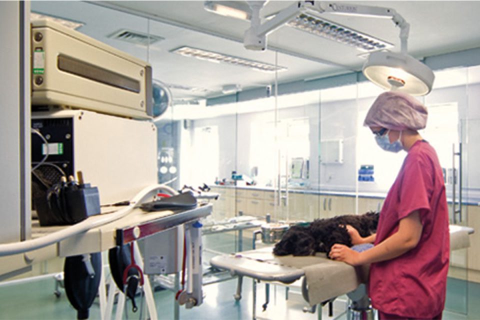
The ability to see inside the body can speed up a diagnosis or hugely increase our understanding of what your pet is suffering from. Golden Valley Vets is unique in the area in the range of imaging equipment we have for a first opinion practice. All our equipment is on site and available 24 hours a day – we do not have to wait for someone to visit or a large lorry to turn up with a scanner in it.
Golden Valley therefore offers a huge advantage to you with fast cost effect imaging available instantly. We may, for example, need to look for a change or injury to a bone, the condition of the heart or lungs, clarification of any issues with the abdomen, or investigating symptoms relating to the spine or the brain.
Sometimes several different imaging techniques need to be combined to put together a complete picture of the area of the body being investigated but this can now all be done with one visit to our hospital.
MRI scanner
There are some situations where X Rays, either standard or CT, are not the best technique as both rely on differences in tissue density to produce an image.
The most common areas where this applies are brain and spinal cord where pathological changes are often not detectable by radiographic means. Some joint and tendon problems can also fall into this category as cartilage in a joint for example is difficult to see using X rays.
An MRI does not use X rays but creates an image from radio waves released from the tissue when a radio frequency pulse is applied and then turned off within a powerful magnetic field. The physics of this are complex but the final result is to detect tissues with differing water content which again can be manipulated digitally to produce a viewable and useful image. We use a rare earth (gadolinium) based intravenous contrast material to enhance areas of high blood flow or inflammation in the images.
MRI is particularly useful for detecting changes in brain and spinal cord and assessing intervertebral discs.
We have a VetMRI scanner (Magnetic Resonance Imaging)

Ultrasound
These are the situations where we use our Ultrasound to view the area we are investigating. Ultrasound works by producing a very high frequency sound wave from a probe which is applied to the body. The sound pulses are reflected from the different tissues and are detected by the same probe. A computer then analyses the received pulses to create a representation of the tissues under investigation. This has the advantage or being dynamic so is invaluable for studying a moving object such as the heart where the movement of valves, thickness of the heart wall and the movement of blood within the heart and vessels can all be studied.
Ultrasound is also invaluable for looking at fluid filled structures such as urinary bladder or gall bladder. It is a quick and easy technique for finding abdominal masses or looking at the structure of kidneys, liver or spleen for example. It can be used for measuring the thickness of intestinal walls and looking at the movement of the gut. It is safe for animal and operator, non-invasive and can usually be performed conscious or under light sedation.
X-rays
A traditional X ray is a two dimensional grey scale image showing the different densities of the tissues through which the X rays have travelled. It is a very useful technique particularly as we are all very familiar with the appearance of radiographs taken with standard views. It is very good for showing changes in dense tissues such as bone and is useful for showing the sizes and positions of different organs where there are differences between the densities of adjacent tissues.
The disadvantage of plain X rays is that many organs are of similar density so it can be difficult to distinguish between them or detect small changes which may still be clinically significant. The two-dimensional nature of the image also means that different areas of the body are ‘overlaid’ on each other which again can make interpretation challenging. For this reason, we usually take at least two images at ninety degree angles to obtain as much information as possible.